File:Chordoma.PNG: Difference between revisions

From Citizendium
Jump to navigation Jump to search
imported>Daniel Mietchen
(filled in)
(== Summary == Importing file)
Tag: Server-side upload
 
(One intermediate revision by one other user not shown)
Line 1: Line 1:
== Summary ==
== Summary ==
{{Image_Details
Importing file
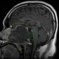
|description  = [[MRI|MRI scan]] of a patient with a very pronounced [[clivus|clival]] [[chordoma]], showing "a large clival based [[tumour]] with involvement of the [[Dura mater|dura]] at the region of the whole clivus where the tumour had compressed the [[brain stem]] anteriorly". The yellow circle highlights the tumour, the green one the clival region.
|author      = [[CZ:Ref:Hassan 2008 Combined use of maxillomandibular swing approach and neurosurgical ultrasonic aspirator in the management of extensive clival chordoma: a case report|Hassan et al., 2008]], with circular markers added by [[User:D. Matt Innis|Matt Innis]]
|copyright    = [[CZ:Ref:Hassan 2008 Combined use of maxillomandibular swing approach and neurosurgical ultrasonic aspirator in the management of extensive clival chordoma: a case report|Hassan et al., 2008]]
|source      = Fig. 1 of [[CZ:Ref:Hassan 2008 Combined use of maxillomandibular swing approach and neurosurgical ultrasonic aspirator in the management of extensive clival chordoma: a case report|Hassan et al., 2008]]
|date-created = 14 August 2007
|pub-country  = United States
|notes        =
|versions    =
}}
 
== Licensing/Copyright status ==
{{CC|by|2.0}}

Latest revision as of 19:52, 11 March 2022

Summary

Importing file

File history

Click on a date/time to view the file as it appeared at that time.

Date/TimeThumbnailDimensionsUserComment
current19:52, 11 March 2022Thumbnail for version as of 19:52, 11 March 2022639 × 639 (348 KB)Maintenance script (talk | contribs)== Summary == Importing file

The following 2 pages use this file: