File:Blood-brain barrier and proinflammatory cytokines.png: Difference between revisions
imported>Daniel Mietchen ({{Image_Details |description = to follow |author = |copyright = |source = [http://www.jneuroinflammation.com/content/1/1/22 http://www.jneuroinflammation.com/content/1/1/22] |date-created = |pub-country = |notes = |versions = }}) |
imported>Daniel Mietchen |
||
| Line 1: | Line 1: | ||
== Summary == | == Summary == | ||
{{Image_Details | {{Image_Details | ||
|description = to | |description = Schematic drawing of [[cellular signalling|cellular regulation]] of [[extracellular]] [[glutamate]] [[concentration (chemistry)|concentrations]] ([Glu]ec) in the presence of the [[proinflammatory]] [[cytokine]]s [[tumor necrosis factor-α]] (TNF-α), [[interleukin-1β|interleukin (IL)-1β]], and [[interleukin-6|IL-6]]. Possible pathophysiology underlying mental fatigue at the cellular level is outlined below. TNF-α, IL-1β and IL-6 attenuate [[astroglia]]l [[glutamate uptake transport]] and disintegrate the [[blood-brain barrier]], allowing [[glutamate]] from the [[blood]] to enter the [[brain]]. The overall result is slightly increased [Glu]ec. Tumor necrosis factor-alfa also decreases oligodendroglial cell glutamate uptake [78], while microglial glutamate uptake has been demonstrated to increase (Persson, M., Hansson, E., and Rönnbäck, L, to be published), though not to levels to compensate for the decreased astroglial glutamate uptake capacity. Due to increased [Glu]ec, astroglial swelling is shown. Below: Hypothetic cellular events underlying mental fatigue. Slightly increased [Glu]ec could make the glutamate neurotransmission less distinct (decrease the signal-to-noise ratio). At the cellular level, there would be astroglial swelling, which in turn would decrease the local extracellular (ec) volume and, as a consequence, lead to further increased [Glu]ec. Astroglial swelling also depolarizes the astroglial cell membrane, which further attenuates the electrogenic glutamate uptake and, in addition, the astroglial K+ uptake capacity. As a consequence, even [K+]ec may rise. The increased [K+]ec, together with decreased glutamine production and reduced glucose uptake concomitant with the decreased glutamate uptake, could lead to decreased presynaptic glutamate release and thereby decreased glutamate transmission, which, according to our hypothesis, is one cellular correlate to mental fatigue/exhaustion. Increased extracellular glutamate levels in the prefrontal region could lead to inhibition of the brain stem nuclei locus coeruleus (LC) and raphe nuclei and thereby inhibit noradrenaline (NA) and serotonin (5-HT) release in the cerebral cortex resulting in decreased astroglial metabolism and neuronal metabolic supply. Increased neuronal excitability may be part of the loudness and light sensitivity often accompanying the mental fatigue. In addition, the decrease in noradrenaline and serotonin release might be part of decreased attention and the appearance of depression often accompanying the mental fatigue. | ||
|author = | |author = [[CZ:Ref:Rönnbäck 2004 On the potential role of glutamate transport in mental fatigue|Rönnbäck and Hansson, 2004]] | ||
|copyright = | |copyright = [[CZ:Ref:Rönnbäck 2004 On the potential role of glutamate transport in mental fatigue|Rönnbäck and Hansson, 2004]] | ||
|source = | |source = Part of Fig. 1 from {{CZ:Ref:Rönnbäck 2004 On the potential role of glutamate transport in mental fatigue}} | ||
|date-created = | |date-created = 30 August 2004 | ||
|pub-country = | |pub-country = United States | ||
|notes = | |notes = | ||
|versions = | |versions = [[:Blood-brain barrier schematic.png]] | ||
}} | }} | ||
Schematic drawing of cellular regulation of extracellular glutamate concentrations ([Glu]ec) in the presence of the proinflammatory cytokines tumor necrosis factor-α (TNF-α), interleukin (IL)-1β, and IL-6. Possible pathophysiology underlying mental fatigue at the cellular level is outlined below. TNF-α, IL-1β and IL-6 attenuate astroglial glutamate uptake transport and disintegrate the BBB, allowing glutamate from the blood to enter the brain. The overall result is slightly increased [Glu]ec. Tumor necrosis factor-alfa also decreases oligodendroglial cell glutamate uptake [78], while microglial glutamate uptake has been demonstrated to increase (Persson, M., Hansson, E., and Rönnbäck, L, to be published), though not to levels to compensate for the decreased astroglial glutamate uptake capacity. Due to increased [Glu]ec, astroglial swelling is shown. Below: Hypothetic cellular events underlying mental fatigue. Slightly increased [Glu]ec could make the glutamate neurotransmission less distinct (decrease the signal-to-noise ratio). At the cellular level, there would be astroglial swelling, which in turn would decrease the local extracellular (ec) volume and, as a consequence, lead to further increased [Glu]ec. Astroglial swelling also depolarizes the astroglial cell membrane, which further attenuates the electrogenic glutamate uptake and, in addition, the astroglial K+ uptake capacity. As a consequence, even [K+]ec may rise. The increased [K+]ec, together with decreased glutamine production and reduced glucose uptake concomitant with the decreased glutamate uptake, could lead to decreased presynaptic glutamate release and thereby decreased glutamate transmission, which, according to our hypothesis, is one cellular correlate to mental fatigue/exhaustion. Increased extracellular glutamate levels in the prefrontal region could lead to inhibition of the brain stem nuclei locus coeruleus (LC) and raphe nuclei and thereby inhibit noradrenaline (NA) and serotonin (5-HT) release in the cerebral cortex resulting in decreased astroglial metabolism and neuronal metabolic supply. Increased neuronal excitability may be part of the loudness and light sensitivity often accompanying the mental fatigue. In addition, the decrease in noradrenaline and serotonin release might be part of decreased attention and the appearance of depression often accompanying the mental fatigue. | |||
== Licensing/Copyright status == | == Licensing/Copyright status == | ||
{{CC|by|2.0}} | {{CC|by|2.0}} | ||
Revision as of 09:25, 2 June 2010
Summary
| Title / Description
|
Schematic drawing of cellular regulation of extracellular glutamate concentrations ([Glu]ec) in the presence of the proinflammatory cytokines tumor necrosis factor-α (TNF-α), interleukin (IL)-1β, and IL-6. Possible pathophysiology underlying mental fatigue at the cellular level is outlined below. TNF-α, IL-1β and IL-6 attenuate astroglial glutamate uptake transport and disintegrate the blood-brain barrier, allowing glutamate from the blood to enter the brain. The overall result is slightly increased [Glu]ec. Tumor necrosis factor-alfa also decreases oligodendroglial cell glutamate uptake [78], while microglial glutamate uptake has been demonstrated to increase (Persson, M., Hansson, E., and Rönnbäck, L, to be published), though not to levels to compensate for the decreased astroglial glutamate uptake capacity. Due to increased [Glu]ec, astroglial swelling is shown. Below: Hypothetic cellular events underlying mental fatigue. Slightly increased [Glu]ec could make the glutamate neurotransmission less distinct (decrease the signal-to-noise ratio). At the cellular level, there would be astroglial swelling, which in turn would decrease the local extracellular (ec) volume and, as a consequence, lead to further increased [Glu]ec. Astroglial swelling also depolarizes the astroglial cell membrane, which further attenuates the electrogenic glutamate uptake and, in addition, the astroglial K+ uptake capacity. As a consequence, even [K+]ec may rise. The increased [K+]ec, together with decreased glutamine production and reduced glucose uptake concomitant with the decreased glutamate uptake, could lead to decreased presynaptic glutamate release and thereby decreased glutamate transmission, which, according to our hypothesis, is one cellular correlate to mental fatigue/exhaustion. Increased extracellular glutamate levels in the prefrontal region could lead to inhibition of the brain stem nuclei locus coeruleus (LC) and raphe nuclei and thereby inhibit noradrenaline (NA) and serotonin (5-HT) release in the cerebral cortex resulting in decreased astroglial metabolism and neuronal metabolic supply. Increased neuronal excitability may be part of the loudness and light sensitivity often accompanying the mental fatigue. In addition, the decrease in noradrenaline and serotonin release might be part of decreased attention and the appearance of depression often accompanying the mental fatigue. |
|---|---|
| Author(s)
|
Rönnbäck and Hansson, 2004 |
| Copyright holder
|
Rönnbäck and Hansson, 2004 See below for license/re-use information. |
| Source
|
Part of Fig. 1 from Rönnbäck L, Hansson E (2004). "On the potential role of glutamate transport in mental fatigue". J Neuroinflammation 1 (1): 22. DOI:10.1186/1742-2094-1-22. PMID 15527505. PMC PMC533886. Research Blogging. [e] |
| Date created
|
30 August 2004 |
| Country of first publication
|
United States |
| Notes
|
You can edit this page and add notes here which may be useful to people who wish to re-use this media. |
| Other versions
|
Blood-brain barrier schematic.png |
| Using this image on CZ
|
Copy the code below to add this image to a Citizendium article, changing the size, alignment, and caption as necessary.
|
Please send email to manager A T citizendium.org .
Schematic drawing of cellular regulation of extracellular glutamate concentrations ([Glu]ec) in the presence of the proinflammatory cytokines tumor necrosis factor-α (TNF-α), interleukin (IL)-1β, and IL-6. Possible pathophysiology underlying mental fatigue at the cellular level is outlined below. TNF-α, IL-1β and IL-6 attenuate astroglial glutamate uptake transport and disintegrate the BBB, allowing glutamate from the blood to enter the brain. The overall result is slightly increased [Glu]ec. Tumor necrosis factor-alfa also decreases oligodendroglial cell glutamate uptake [78], while microglial glutamate uptake has been demonstrated to increase (Persson, M., Hansson, E., and Rönnbäck, L, to be published), though not to levels to compensate for the decreased astroglial glutamate uptake capacity. Due to increased [Glu]ec, astroglial swelling is shown. Below: Hypothetic cellular events underlying mental fatigue. Slightly increased [Glu]ec could make the glutamate neurotransmission less distinct (decrease the signal-to-noise ratio). At the cellular level, there would be astroglial swelling, which in turn would decrease the local extracellular (ec) volume and, as a consequence, lead to further increased [Glu]ec. Astroglial swelling also depolarizes the astroglial cell membrane, which further attenuates the electrogenic glutamate uptake and, in addition, the astroglial K+ uptake capacity. As a consequence, even [K+]ec may rise. The increased [K+]ec, together with decreased glutamine production and reduced glucose uptake concomitant with the decreased glutamate uptake, could lead to decreased presynaptic glutamate release and thereby decreased glutamate transmission, which, according to our hypothesis, is one cellular correlate to mental fatigue/exhaustion. Increased extracellular glutamate levels in the prefrontal region could lead to inhibition of the brain stem nuclei locus coeruleus (LC) and raphe nuclei and thereby inhibit noradrenaline (NA) and serotonin (5-HT) release in the cerebral cortex resulting in decreased astroglial metabolism and neuronal metabolic supply. Increased neuronal excitability may be part of the loudness and light sensitivity often accompanying the mental fatigue. In addition, the decrease in noradrenaline and serotonin release might be part of decreased attention and the appearance of depression often accompanying the mental fatigue.
Licensing/Copyright status
This media, Blood-brain barrier and proinflammatory cytokines.png, is licenced under the Creative Commons Attribution 2.0 Unported License
You are free:
To Share — To copy, distribute and transmit the work; To Remix — To adapt the work.
Under the following conditions:
Attribution — You must attribute the work in the manner specified by the author or licensor (but not in any way that suggests that they endorse you or your use of the work).
For any reuse or distribution, you must make clear to others the licence terms of this work (the best way to do this is with a link to this licence's web page). Any of the above conditions can be waived if you get permission from the copyright holder. Nothing in this licence impairs or restricts the author's moral rights.
Read the full licence.
File history
Click on a date/time to view the file as it appeared at that time.
| Date/Time | Thumbnail | Dimensions | User | Comment | |
|---|---|---|---|---|---|
| current | 19:52, 11 March 2022 | 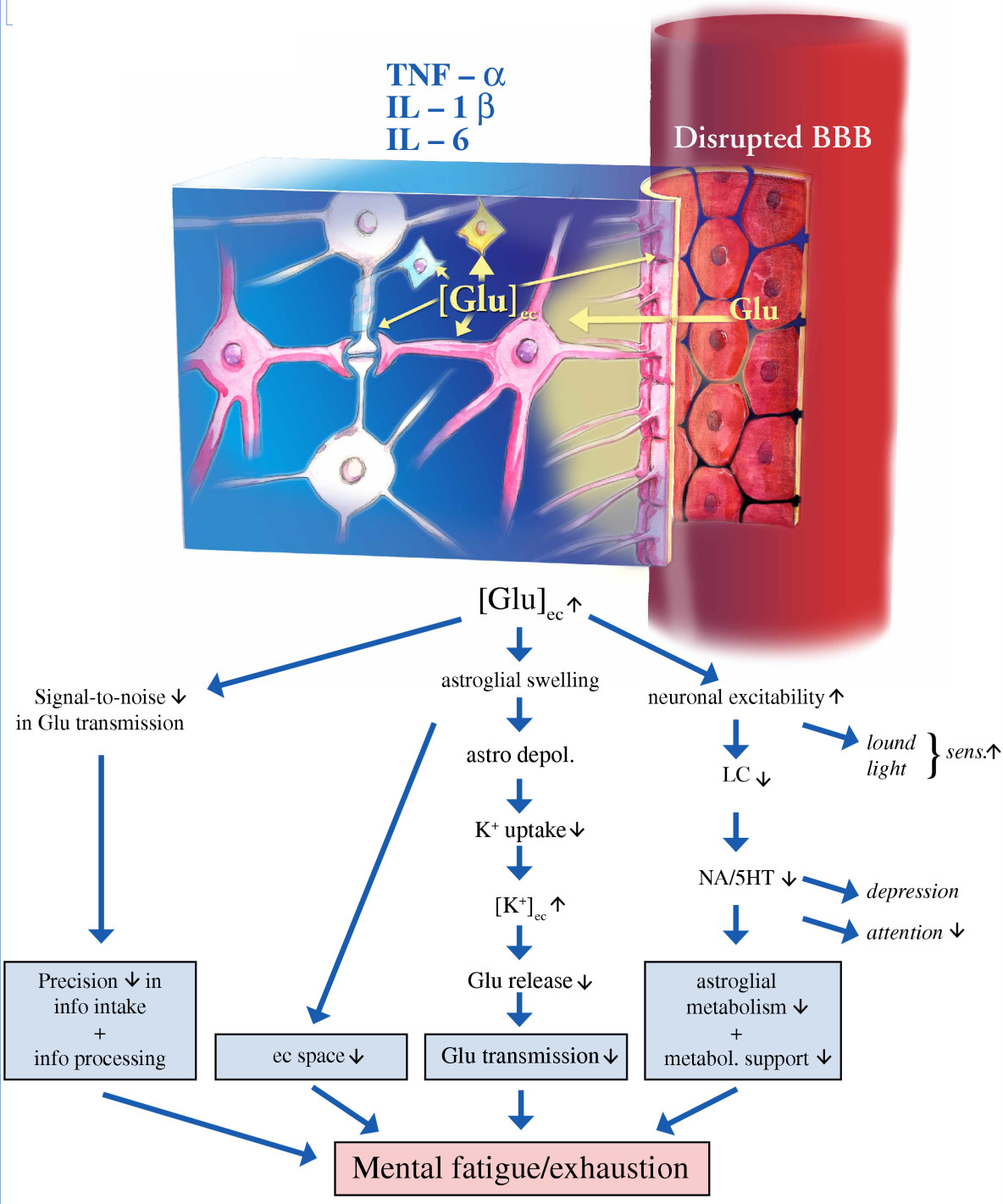 | 1,184 × 1,421 (957 KB) | Maintenance script (talk | contribs) | == Summary == Importing file |
You cannot overwrite this file.
File usage
There are no pages that use this file.