Klebsiella pneumoniae: Difference between revisions
imported>Hirenkumar B Patel No edit summary |
imported>Hirenkumar B Patel |
||
| Line 23: | Line 23: | ||
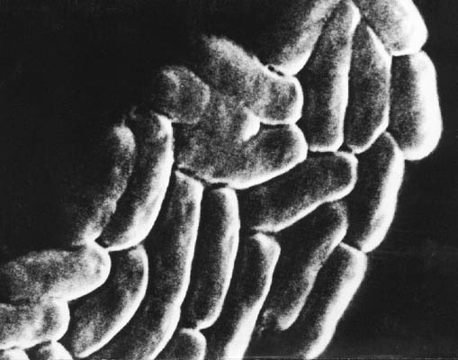
== Image of Klebsiella pneumoniae == | == Image of Klebsiella pneumoniae == | ||
[[Image:Klebsiella pneumoniae.jpg]] Image is from Microbiologia! | [[Image:Klebsiella pneumoniae.jpg]] Image is from Microbiologia! | ||
==Description and significance== | ==Description and significance== | ||
Revision as of 20:32, 30 April 2009
For the course duration, the article is closed to outside editing. Of course you can always leave comments on the discussion page. The anticipated date of course completion is May 21, 2009. One month after that date at the latest, this notice shall be removed. Besides, many other Citizendium articles welcome your collaboration! |
Classification
| Klebsiella pnemoniae | ||||||||||||||
|---|---|---|---|---|---|---|---|---|---|---|---|---|---|---|
 | ||||||||||||||
| Scientific classification | ||||||||||||||
| ||||||||||||||
| Binomial name | ||||||||||||||
| Pseudomonas putida |
Klebsiella pnemoniae is a species of Genus: Klebsiella. The Family: Enterobacteriaceae. The Order: Enterobacteriales. The Class: Gammaproteobacteria. The Phylum: Proteobacteria. and The Domain: Bacteria(1).
Image of Klebsiella pneumoniae
Description and significance
K. pneumoniae is a gram negative bacteria. It is faculative aerobic, meaning it is has characteristic features of becoming aerobic and anaerobic. It is rod shaped and its dimensions are 2 X 0.5 micro meter. K. pneumoniae is also known as nasty bacteria. Klebsiella (pneumoniae) is commonly found in bacteria directed pneumoniae patients. Friedlancer C. Uber was the first person to discover that Klebsiella is a pathogenic bacteria that causes pneumoniae (2). The 160nm thick fine fiber capsule makes the bacteria pathogenic (3)[1]. This bacteria is found in different places in human body such as gastrointestinal tract and nasopharynx. (4) Naturally it is found in the soil, water, vegetables and sewage.
Genome structure
There are lots of research being done on sequencing the genome. Some places such as (Genome Sequencing Center)at Washington University in St. Louis and (PLOS genetics). At Genome Sequencing Center the genome was named Klebsiella pneumoniae subsp. pneumoniae MGH 78578 ATCC 700721 [2] It was isolated from 66 year old male patient in 1994. It contained one chromosome of 5.3Mbp. There are plasmids pKPN3, pKPN4, pKPN5, pKPN6 and pKPN7 the plasmid length afre respectively 0.18, 0.11, 0.089, 0.0043, and 0.0035 Mbp. Similarly, at PLOS genetics Kp342 was studied. It contained one chromose of 5,641,239 bp. There are two plasmids pKP187 of length 182,922 bp. and pKP91 of length 91,096 bp (5)[3]. The whole genome is sequenced. At both places the DNA was circular.
Cell structure and metabolism
Cell Structure
Klebsiella pneumoniae contains Lipopolysaccharide and capsular polysaccharide capsule around the cell. Capsular polysaccharide are responsible for part of ribosomal immunoprotective activity. It also protects bacteria from phagocytosis. Klebsiella pneumoniae has memberanous vesicles that containe lipopolysaccharide, proteins, and lipids which help capsular polysaccharide with immunoprotective activity(6)[4].
Metabolism
Klebsiella pneumoniae fixes nitrogen from atmosphere. It synthesized amino acids from nitrogen. It utilizes nitriles as nitrogen sources(7)[5]. In the study of the genes of Klebsiella pneumoniae it was found that glnA and glnG regulates the nitrogen fixing activities(8).
Ecology
Klebsiella pneumoniae is found in water, sewage, in animals and mammals, in short it could be found anywhere in the ecological environment. It is pathogenic bacteria. The study on Klebsiella pneumoniae is found every where due to diseases caused by it. It affects almost all of the organs in the body.
Pathology
We all have millions of bacteria in our gastrointestinal tracts, primarily in the colon. Klebsiella pneumoniae is also in the gut. But once if it gets out it causes serious infections. The infections tend to occur in people with weak immune system. So Klebsiella infections can be deadly for people with immunodeficiency.
References
(1)NCBI, [6]
(2)Friedlander C. Uber die scizomyceten bei der acuten fibrosen pneumonie. Arch Pathol Anat Physiol Klin Med 1882. 87:319-24
(3)Amako, K., Meno, Y., and Takade, A. "Fine Structures of the Capsules of Klebsiella pneumoniae and Escherichia coli K1". Journal of Bacteriology. 1988. Volume 170, No. 10. p. 4960-4962[7]
(4)Casewell M, Talsania H G. Predominance of certain klebsiella capsular types in hospitals in the United Kingdom. J Infect. 1979;1:77–79.
(5)Derrick E. Fouts, Heather L. Tyler, Robert T. DeBoy, Sean Daugherty, Qinghu Ren, Jonathan H. Badger, Anthony S. Durkin, Heather Huot, Susmita Shrivastava, Sagar Kothari, Robert J. Dodson, Yasmin Mohamoud, Hoda Khouri, Luiz F. W. Roesch, Karen A. Krogfelt, Carsten Struve, Eric W. Triplett, Barbara A. Methe. "Complete Genome Sequence of the N2-Fixing Broad Host Range Endophyte Klebsiella pneumoniae 342 and Virulence Predictions Verified in Mice".
(6)Jean-Michel Fournier, Colette Jolivet-Reynaud, Marie-Madeleine Riottot, and Hélène Jouin. "Murine Immunoprotective Activity of Klebsiella pneumoniae Cell Surface Preparations: Comparative Study with Ribosomal Preparations", Infect Immun. 1981 May; 32(2): 420–426. PMCID: PMC351459
(7)Cerniglia, Carl E., Heinze, Thomas M., Nawaz, Mohamed S. "Metabolism of benzonitrile and butyronitrile by Klebsiella pneumoniae", 1992 [8]
(8)Espin G, Alvarez-Morales A, Merrick M. "Complementation analysis of glnA-linked mutations which affect nitrogen fixation in Klebsiella pneumoniae". Mol Gen Genet. 1981. Volume 184, No. 2. p. 213-7. [9]
