Thoracic wall: Difference between revisions
Jump to navigation
Jump to search

imported>Robert Badgett (New page: {{subpages}} right|thumb|350px|{{#ifexist:Template:Gray-image112.gif/credit|{{Gray-image112.gif/credit}}<br/>|}}The rib cage viewed from the front. The thoracic...) |
imported>Meg Taylor No edit summary |
||
| (2 intermediate revisions by 2 users not shown) | |||
| Line 1: | Line 1: | ||
{{subpages}} | {{subpages}} | ||
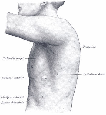
{{Image|Gray-image1215.gif|right|350px|The surface of the left side of the thorax.}} | |||
The thoracic wall is "the outer margins of the thorax containing skin, deep fascia; thoracic vertebrae; ribs; sternum; and muscles."<ref>{{MeSH}}</ref> | The thoracic wall is "the outer margins of the thorax containing skin, deep fascia; thoracic vertebrae; ribs; sternum; and muscles."<ref>{{MeSH}}</ref> | ||
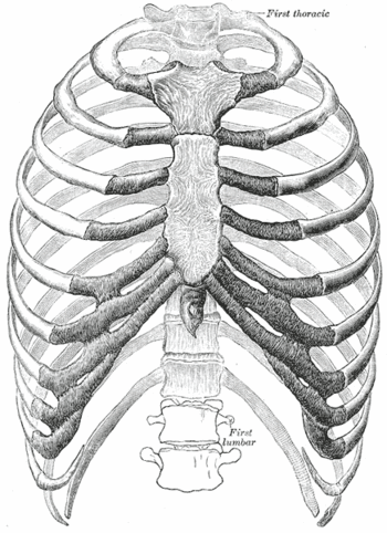
{{Image|Gray-image112.gif|right|350px|The rib cage viewed from the front.}} | |||
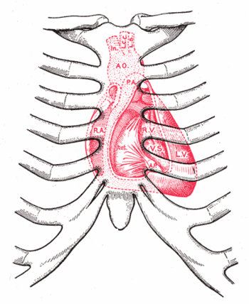
{{Image|Gray-image1218.gif|right|350px|Diagram showing relations of opened heart to front of thoracic wall.}} | |||
==References== | ==References== | ||
{{reflist}} | |||
Latest revision as of 03:12, 7 October 2013
The thoracic wall is "the outer margins of the thorax containing skin, deep fascia; thoracic vertebrae; ribs; sternum; and muscles."[1]
References
- ↑ Anonymous (2024), Thoracic wall (English). Medical Subject Headings. U.S. National Library of Medicine.


