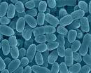
Bordetella pertussis
| ;"|Bordetella pertussis | ||||||||||||||
|---|---|---|---|---|---|---|---|---|---|---|---|---|---|---|
| ;" | Scientific classification | ||||||||||||||
| ||||||||||||||
| Binomial name | ||||||||||||||
| Bordetella pertussis |
Description and Significance
Bordetella pertussis, commonly known as whooping cough, was first defined in the 16th century. It is a respiratory tract infection depicted by a paroxysmal cough. B. pertussis is extremely tiny, and is a Gram-negative aerobic coccobacillus. It can appear in singles or pairs. Before vaccinations were prevalent, B. pertussis was a major cause of death among children and infants. After the pertussis vaccine was introduced, reported cases of this infection decreased by more than 99%. Even though this infection has been contained for the most part, it is still remains a disease that is of major concern. [1]
Ecology
Humans are the only home for B. pertussis. The pathogen is contagious and can be transferred from person to person through aerosolized droplets by sneezing or coughing. This Gram-negative pleomorphic bacillus attaches to and damages ciliated respiratory epithelium. Its main residence is within the trachea and the bronchi. The pathogen will cease to exist in the environment if it is not embedded in the respiratory mucus of the host. [2]
Genome Structure
Tomaha I, a strain of B. pertussis has its genome completely sequenced. It consists of one circular chromosome containing 4.086,189 nucleotides. GC bonds makes up approximately 67% of the genome. The coding density is 82%.
Cell Structure and Metabolism
B. pertussis uses aerobic respiration as its metabolism. Its cell structure consists of an inner membrane, outer membrane, and periplasmic space. The periplasmic space contains a thin peptidoglycan wall layer in between it. Lipopolysaccharides reside on the outer membrane. These types of endotoxins are not seen in any other Gram-negative bacteria. The lipopolysaccharides in B. pertussis contain two forms, which differ in their phosphate composition of the lipid region of the lipopolysaccharide. Lipid X is the designation of this unusual lipid, which is usually in the Lipid A form. Lipid X's function is not known. [2]
Pathology
Humans are the only host for B. pertussis. The respiratory disease caused by B. pertussis is known as pertussis. It is highly contagious, especially in the early stages, and consists of violent coughing which is immediately followed by "whooping" sounds during the intake or air. Hence, this disease is also known as "Whooping Cough". Vomiting is also a symptom of this disease. It also consists of discharge, which contains a sticky mucus. Other symptoms of pertussis are runny nose, coughing sneezing, and a minor fever.
The whooping cough is due to one of the virulence factors of B. pertussis, adenylate cyclase toxin. (CyaA) CyaA is a single polypeptide which attacks eukaryotic cells by directly moving across the plasma membrane of the target cell. It helps in the initiation of the infection by reducing phagocytic activity. CyaA contains two domains. An enzymatic domain and a binding domain, which attaches itself to the surface of the host cells. CyaA is only active when calmodulin, a eukaryotic regulatory molecule, is present. Therefore it is not present in prokaryotes. Pertussis toxin is another major virulence factor of B. pertussis. It is a major factor of the respiratory infection, especially in the early stages. Pertussis toxins' main target are the macrophages in the respiratory tract. By doing so, pertussis toxin is able to promote the infection. Another virulence factor B. pertussis that is responsible for cell adhesion to host cells is filamentous hemagglutinin. [3]
Frequency
B. pertussis occurs mostly during the summer months, usually occurring between June and September. Lifelong immunity is not attained, even if an individual is vaccinated or has even acquired the disease once before. The vaccination usually lasts about three to five years. B. pertussis infects people of every age, but infants are more susceptible than any other group. [1]
Colonization
Two of the main toxins of B. pertussis that are responsible for colonization are filamentous hemagglutinin and pertussis toxin. Galactose resides on a sulfated glycolipid known as sulfatide is the region where filamentous hemagglutinin binds. These sulfatides are primarily located on the surface of ciliated cells. Any sort of mutation that occurs to the filamentous hemagglutinin structural gene causes a decrease in the organisms ability to colonize.
Pertussis toxin also aids in colonization. This toxin is secreted into two main areas. These two areas are the extracellular fluid and the cell. It is responsible for attachment to the tracheal epithelium. It has six components. S1, S2, S3, two S4's, S5, and S6. S2 and S3 are responsible for binding the bacteria to the host cells. S2 binds to glycolipids located on the ciliated epithelial cells and S3 binds to glycoproteins of phagocytic cells. The main glycolipid that S2 binds to is known as lactosylceramide. There are effective antibodies that prevent pertussis toxin from colonizing ciliated cells. This helps to fight against the infection. It is believed that filamentous hemagglutinin binds to phagocytic surfaces through S3 subunit to facilitate its own engulfment into the cell. The reason for this is not known. One reason might be to allow B. pertussis to enter the cell and become an intracellular parasite. [2]
Application to Biotechnology
To fight the fatal disease known as whooping cough, B. pertussis has been used in the medical field to aid in finding a vaccine. The vaccine that is currently being utilized has reduced the occurrence of the disease dramatically. The vaccine uses a chemically-inactivated whole cell vaccine. Side effects are definitely a factor, but the effects do not outweigh the risks of the disease. Examples of some side effects are acute encephalopathy, convulsions and hypotensive-hyporesponsive episodes Currently, a new vaccine is being developed which utilizes recombinant DNA technology. [2]
Diagnosis
Pertussis is suggested when patients have:[1]
- Posttussive emesis
- Inspiratory whoop
- Paroxysmal cough
Current Research
A major factor in most bacterial infections is the ability of the microbe to attach to the hosts cells. B. pertussis' ability to attach to and colonize in the hosts cells is investigated using different antibodies. Two classes of antibodies are known to effectively reduce attachment to epithelial host cells. They are known as IgG and IgA. Fimbriae-specific antibodies help with accomplishing this feat. Many other antibodies are available, but none of them are effective in preventing the bacterial cells from attaching to the host cells. The only exception is antifilamentous hemaglutinin antibodies. Even though it is effective, the ability of antifilamentous hemaglutinin antibodies to prevent attachment to cells is extremely small compared to that of fimbriae-specific antibodies.
Antigenic divergence was a vaccine that was very effective in combating against pathogenic microbe organisms such as B. pertussis. This will no longer be so. Despite heavy vaccinations, the occurrence of B. pertussis keeps resurfacing due to the fact that there is an antigenic divergence between the vaccine strains and the circulating strains. In the early 1960's and 1970's, the two alleles that were dominant in prevaccination strains were the ptxa2 allele and the ptxa4 allele. This changed in the early 1970's, and the allele that was expressed most frequently was the ptxa1 allele, which is still the case today. Allele prn1 was also the more dominant allele in prevaccination tests. This did not hold over time and two new alleles became dominant, prn2 and prn3. Due to these shifts in allele frequencies, vaccines that were effective in earlier years are no longer effective today. They are not able to combat the new alleles that have become more dominant. New vaccines have to be developed to help fight these newly dominant strains of B. pertussis.
New research has been initiated to help find a new effective vaccination forB. pertussis. The current vaccine is known as a whole-cell vaccine, meaning it utilizes whole dead B. pertussis cells. This vaccine has known to be very effective. Very few people have developed an allergic reaction against this vaccine though. The reason for this is due the bacteria's lipopolysaccharides endotoxic activity. These lipopolysaccharides are found on the outer membrane in all Gram-negative bacteria. Lipopolysaccharides contain a toxic region, which is located in the lipid region. These lipids are located inside the membrane of the bacteria. Lipopolysaccharides are sometimes released in to the environment when the bacteria dies. This leads to the toxic regions of the lipid region of the lipopolysaccharide to be exposed. PagP and PagL are two enzymes that contain lipopolysaccharide modifying agents. PagP increases the endotoxic activity of the lipopolysaccharide and the whole bacterial cell while PagL decreased endotoxic activity of the lipopolysaccharide, but increased endotoxin activity of the whole bacterial cells. The reason for the increase in endotoxin activity in whole bacterial cells, even in the presence of PagL is a consequence of the increase in lipopolysaccharide activity into the cell overall. [2]
References
- ↑ Cornia PB, Hersh AL, Lipsky BA, Newman TB, Gonzales R (2010). "Does this coughing adolescent or adult patient have pertussis?". JAMA 304 (8): 890-6. DOI:10.1001/jama.2010.1181. PMID 20736473. Research Blogging.
[1]↑ http://emedicine.medscape/article/967268
[2]↑ http://microbewiki.kenyon.edu/index.php/Bordetella_pertussis

